Pelvic Anatomy Ligaments / Pelvis Skeleton Model Female 6 Part 3b Scientific 1000288 H20 4 : Bones and ligaments of the female pelvis.
byAdmin•
0
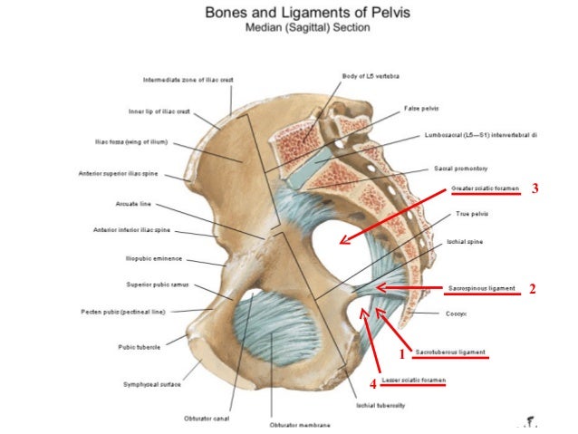
Pelvic Anatomy Ligaments / Pelvis Skeleton Model Female 6 Part 3b Scientific 1000288 H20 4 : Bones and ligaments of the female pelvis.. The pelvis itself is a paired composite structure made up by three bones (ilium, ischium and pubis) that articulates with the sacral part of the axial spine. You can click the image to magnify if you cannot see clearly. It extends to both sides of the pelvic wall. There are ligaments between the sacrum and the ilium, which are called sacroiliac ligaments. Joints and ligaments of the pelvis (anterior view)
The pubocervical ligaments are a pair of fibrous bands that attach the anterior portion of the cervix to the posterior pubic symphysis. There are two major groups of ligaments that provide nearly all the structure of the pelvis. This image shows the posterior back view of the female pelvic brim (the bones and ligaments that forms the pelvic region in the female) showing: It is usually divided into two separate anatomic regions: Über 7 millionen englischsprachige bücher.
Pelvis And Hip Joint Amboss from media-us.amboss.com The pelvis consists of two innominate bones and the sacrum to which coccyx is attached. The cardinal ligament is a paired thickening of the parametrium and pelvic fascia at the base of the broad ligament, which extends between the cervix and vaginal fornix medially to the sidewall of pelvis laterally. The named ligaments of the pelvis mostly arise from the sacrum and attach to varying segments of the pelvic bone. (2017, elsevier) should be consulted. The outlet is formed by the pubic arch, ischial spines, sacrotuberous ligaments, and the coccyx. The ilium, ischium and the pubic bone. It is usually divided into two separate anatomic regions: Über 7 millionen englischsprachige bücher.
The pectineal ligament is strong, and holds suture well.
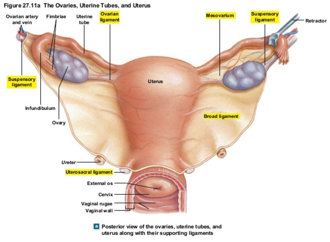
You can click the image to magnify if you cannot see clearly. The broad ligament can be further divided into three components. Thank you for visit anatomynote.com. Other ligaments attached to bony pelvis include the sacrococcygeal ligaments, pubic symphysis ligaments, and endopelvic fascia ligament. There are ligaments between the sacrum and the ilium, which are called sacroiliac ligaments. The pelvic girdle, also known as the hip bone, is composed of three fused bones: The broad ligament is a flat sheet of peritoneum, associated with the uterus, fallopian tubes and ovaries. The pelvic inlet involves three of the four units of which the bone pelvis is composed. We hope you can get the exact information you. It is usually divided into two separate anatomic regions: The pelvis consists of two innominate bones and the sacrum to which coccyx is attached. The inlet to the pelvic canal is at the level of the sacral promontory and superior aspect of the pubic bones. The pelvis is the lower portion of the trunk, located between the abdomen and the lower limbs.
As with the muscles, it is also helpful to know the ligaments of the lumbar. It extends to both sides of the pelvic wall. The broad ligament can be further divided into three components. The pelvis is the lower portion of the trunk, located between the abdomen and the lower limbs. Learning pelvic anatomy is composed of learning bones, muscles, ligaments, nerves and vascular supply.
Clinical Anatomy Of Pelvis from image.slidesharecdn.com The femoral ligaments act to stabilize the ball and socket joint of the hip, connecting to the ilium and the ischium. The cardinal ligaments, also known as the transverse cervical ligaments, the lateral cervical ligaments, or mackenrodt's ligaments, are fibrous bands that attached the cervix to the lateral pelvic walls. The broad ligament is a flat sheet of peritoneum, associated with the uterus, fallopian tubes and ovaries. Bones and ligaments of the female pelvis. The pelvic inlet involves three of the four units of which the bone pelvis is composed. Learning pelvic anatomy is composed of learning bones, muscles, ligaments, nerves and vascular supply. Inner surface of obturator membrane lateral attachment: The named ligaments of the pelvis mostly arise from the sacrum and attach to varying segments of the pelvic bone.
How to use the anatomical labels
The pelvic brim involves the first sacral segment, the iliac and pubis portion, but not the ischium. The pectineal ligament is strong, and holds suture well. Inherent stability of the pelvis is provided by ligaments. A full understanding of pelvic anatomy is required to treat pelvic fractures, to prevent iatrogenic injuries, and to provide the best results. (2017, elsevier) should be consulted. Additional ligaments may be found in the female pelvis. The uterosacral ligaments were the most rigid whether at low or high deformation, while the round ligament was more rigid than the broad ligament. Cardinal ligament and the uterosacral ligaments provide apical support for the uterus and upper vagina. citation needed this facilitates reconstruction of the floor of the inguinal canal. Bones and ligaments of the female pelvis. Ligaments of the lumbar spine and pelvis. Pelvic bone and ligaments anatomy. The pelvic inlet involves three of the four units of which the bone pelvis is composed.
The pelvis's frame is made up of the bones of the pelvis, which connect the axial skeleton to the femurs, and therefore acts in weight bearing of the upper body. The pubocervical ligaments are a pair of fibrous bands that attach the anterior portion of the cervix to the posterior pubic symphysis. The pelvic brim involves the first sacral segment, the iliac and pubis portion, but not the ischium. Bones and ligaments of the female pelvis. citation needed this facilitates reconstruction of the floor of the inguinal canal.
Uterus Anatomy Ligaments Anatomy Drawing Diagram from d1yboe6750e2cu.cloudfront.net Anatomy of the female pelvis : Inner surface of obturator membrane lateral attachment: How to use the anatomical labels You can click the image to magnify if you cannot see clearly. The pelvic brim involves the first sacral segment, the iliac and pubis portion, but not the ischium. The pectineal ligament is strong, and holds suture well. Bones and ligaments of the female pelvis. Iliolumbar, sacrotuberous and sacrospinous ligaments.
Inner surface of obturator membrane lateral attachment:
The femoral ligaments act to stabilize the ball and socket joint of the hip, connecting to the ilium and the ischium. Bones and ligaments of the female pelvis. As with the muscles, it is also helpful to know the ligaments of the lumbar. A full understanding of pelvic anatomy is required to treat pelvic fractures, to prevent iatrogenic injuries, and to provide the best results. The enclosed space between the inlet and outlet is called the true pelvis, with the plane of the inlet being at right angles to the plane of the outlet. These ligaments firmly hold together the two pubic bones and, consequently, the two innominate bones. The pectineal ligament is usually around 6 cm long in adults. It extends from the lateral pelvic walls on both sides, and folds over the internal female genitalia, covering their surface anteriorly and posteriorly. Cardinal ligament and the uterosacral ligaments provide apical support for the uterus and upper vagina. The outlet is formed by the pubic arch, ischial spines, sacrotuberous ligaments, and the coccyx. There are two major groups of ligaments that provide nearly all the structure of the pelvis. It extends to both sides of the pelvic wall. The pelvic brim involves the first sacral segment, the iliac and pubis portion, but not the ischium.
Ligaments connect one bone to another and provide important stability pelvic anatomy. Anatomy of the human female pelvis: