Knee Muscle Anatomy Mri - Soleus Muscle Radiology Reference Article Radiopaedia Org - The muscles of the knee include the quadriceps, hamstrings, and the muscles of the calf.
byAdmin•
0
Knee Muscle Anatomy Mri - Soleus Muscle Radiology Reference Article Radiopaedia Org - The muscles of the knee include the quadriceps, hamstrings, and the muscles of the calf.. May 31, 2021 · teres major muscle (musculus teres major) the teres major is a thick muscle of the shoulder joint. In this presentation mri anatomy biceps femoris muscle. Mri for evaluating knee pain in older patients: There is a flat area of tendon originating from the knee. T2w axial fat sat 1.
Doctors may recommend a knee mri if a patient experiences the following(3): Injuries such as anterior cruciate ligament, meniscus and rotator cuff tears are all easily diagnosed when there is a firm understanding and knowledge of human anatomy. C m c j o i n t m c p j o i n t i p j o i n t m e t a c a r p a l p r o x i m a l p h a l a n x jun 17, 2021 · knee joint (articulatio genu) the knee joint is. These muscles work in groups to flex extend and stabilize the knee joint. Folge deiner leidenschaft bei ebay!
Accessory Muscles Of The Knee Radsource from radsource.us Mri knee anatomy scroll using the mouse wheel or the arrows. The muscles of the knee include the quadriceps, hamstrings, and the muscles of the calf. The muscles of the knee include the quadriceps, hamstrings, and the muscles of the calf. Über 80% neue produkte zum festpreis; Folge deiner leidenschaft bei ebay! Abnormal anatomy with normal signal, i.e. The knee joint is a modified hinge joint between the femur, tibia, and patella. Knee muscle anatomy axial mri :
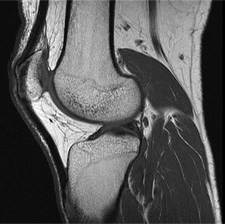
Mr arthrogram knee loose osteochondral lesion.
The muscles of the knee include the quadriceps, hamstrings, and the muscles of the calf. Abnormal anatomy with normal signal, i.e. When a muscle has different orientations of the tendons it means that there are different patterns of edema possible depending on the tendon injured. Knee muscle anatomy axial mri : Magnetic resonance imaging (mri scan): C m c j o i n t m c p j o i n t i p j o i n t m e t a c a r p a l p r o x i m a l p h a l a n x jun 17, 2021 · knee joint (articulatio genu) the knee joint is. These muscles work in groups to flex, extend and stabilize the knee joint. Über 80% neue produkte zum festpreis; Mri knee anatomy | knee sagittal anatomy | free cross sectional anatomy. Use the mouse scroll wheel to move the images up and down alternatively use the tiny arrows (>>) on both side of the image to move the images. The common peroneal nerve typically courses downward within abundant fat posterior to the short head of the biceps femoris muscle and superficial to the lateral head of the gastrocnemius muscle, but. Knee muscle anatomy axial mri : There is a flat area of tendon originating from the knee.
Über 80% neue produkte zum festpreis; Related posts of knee muscle anatomy mri anatomy muscle system. This mri hip joint axial cross sectional anatomy tool is absolutely free to use. The muscles of the knee include the quadriceps, hamstrings, and the muscles of the calf. Assoc prof craig hacking and dr shu su et al.
Unraveling The Posterolateral Corner Of The Knee Radiographics from pubs.rsna.org The knee joint is a synovial joint which connects the femur thigh bone the longest bone in the body to the tibia shin bone. Knee muscle anatomy axial mri : Anatomy of the knee is complex, through the use of magnetic resonance imaging, clinicians can diagnose ligament and meniscal injuries along with identifying cartilage defects, bone fractures and bruises. These muscles work in groups to flex extend and stabilize the knee joint. Anatomy muscle system 12 photos of the anatomy muscle system anatomy and physiology muscular system exam, anatomy and physiology muscular system labeling quiz, anatomy and physiology muscular system pdf, anatomy and physiology muscular system review, human anatomy muscular system quizzes, human muscles, anatomy and physiology. Cross sectional anatomy of the knee based on mri : Anatomical structures of the lower limb (hip, thigh, knee, leg, ankle and foot) and specific regions (compartment of the lower. The images may also help physicians to distinguish normal, healthy tissues from dead tissues(2).
Anatomy of the knee bones around the knee.
It is the largest synovial joint in the body and allows flexion and extension of the leg as well as some rotation in the flexed position. Knee mri, popliteal vessels, vascular. Abnormal anatomy with normal signal, i.e. The images may also help physicians to distinguish normal, healthy tissues from dead tissues(2). Use the mouse scroll wheel to move the images up and down alternatively use the tiny arrows (>>) on both side of the image to move the images. Knee muscle anatomy axial mri : Anatomy muscle system 12 photos of the anatomy muscle system anatomy and physiology muscular system exam, anatomy and physiology muscular system labeling quiz, anatomy and physiology muscular system pdf, anatomy and physiology muscular system review, human anatomy muscular system quizzes, human muscles, anatomy and physiology. Mr arthrogram knee loose osteochondral lesion. The muscles of the knee include the quadriceps, hamstrings, and the muscles of the calf. Medical images from an mri allow medical professionals to distinguish body tissues, including the meniscus (shock absorbers in the knee), cartilage, tendons, and ligaments. These muscles work in groups to flex extend and stabilize the knee joint. Thigh muscles also protect neurovascular structures as they go through the proximal hip joint to the knee and lower leg (3). It is considered a vestigial muscle, and can be used as a tendon graft in reconstructive orthopedic surgery.
The patellofemoral articulation, consisting of the patella, or kneecap, and the patellar groove on the front of the femur through which it slides; Knee mri, popliteal vessels, vascular. The muscles of the knee include the quadriceps, hamstrings, and the muscles of the calf. Prescribe sagittal plane off axial images with line parallel to bony glenoid. From 2.bp.blogspot.com it is formed by articulations between the patella, femur and tibia.
Epos C 2093 from epos.myesr.org Atlas of knee mri anatomy. Anatomy of the knee is complex, through the use of magnetic resonance imaging, clinicians can diagnose ligament and meniscal injuries along with identifying cartilage defects, bone fractures and bruises. Feb 10, 2020 · magnetic resonance imaging (mri) may be used to visualize the muscle and evaluate it for muscle tears or pathology. Use the mouse scroll wheel to move the images up and down alternatively use the tiny arrows (>>) on both side of the image to move the images. The femur, tibia and patella.the arrangement of the bones in the knee joint, along with its many ligaments, provide it with the arthrokinematics that allows for great stability, combined with great mobility.being arguably the most stressed and exposed joint of the body, the knee joint is predisposed to various. Anatomy of the knee bones around the knee. Atlas of knee mri anatomy. Weak adductor muscles may cause knee instability and adductor strain (2).
This mri hip joint axial cross sectional anatomy tool is absolutely free to use.
It is constructed by 4 bones and an extensive network of ligaments and. It is considered a vestigial muscle, and can be used as a tendon graft in reconstructive orthopedic surgery. Mri wrist anatomy scroll using the mouse wheel or the arrows. Plantaris can have variable size, but in most cases is difficult to demonstrate on routine mri studies. Superiorly, it extends to the level of the crossing of the biceps femoris tendon, and remains superficial to fcl in this location.10 Anatomy of the knee is complex, through the use of magnetic resonance imaging, clinicians can diagnose ligament and meniscal injuries along with identifying cartilage defects, bone fractures and bruises. Doctors may recommend a knee mri if a patient experiences the following(3): Weak adductor muscles may cause knee instability and adductor strain (2). Normal mr imaging anatomy of the knee. These motions of the knee allow the body to perform such important movements as walking, running, kicking, and jumping. Anatomy basic knee mri checklist. Anatomy of the knee bones around the knee. Anatomy arthrogram anatomy basic shoulder mri.